When you are preparing for a dental implant, the quality of your pre-surgical evaluation has a direct impact on how your procedure goes. Standard dental X-rays produce a flat, two-dimensional image of your teeth and bone, which is useful for routine diagnostics but limited when a complete picture of your jaw structure is needed. Three-dimensional digital imaging closes that gap by revealing bone density, nerve pathways, sinus positions, and other anatomical details that a conventional X-ray can’t provide. For patients preparing to undergo implant treatment, this technology is not an optional upgrade. It is a foundational part of planning a safe and successful procedure.
At Rockland Dental Specialists, our periodontists and implant specialists use advanced 3D digital imaging as a core component of how we evaluate and plan every implant case. Dr. Shalom Mintz, board-certified in periodontics and implantology, along with periodontist Dr. David Peto and our full clinical team, rely on this technology to assess your anatomy thoroughly before any procedure begins. Every case we take on starts with a complete diagnostic picture, because precise planning is what allows us to achieve outcomes that are both safe and lasting. If you are considering dental implants, understanding the imaging process is one of the most important steps in knowing what to expect from your care.
How 3D Digital Imaging Works in a Dental Setting
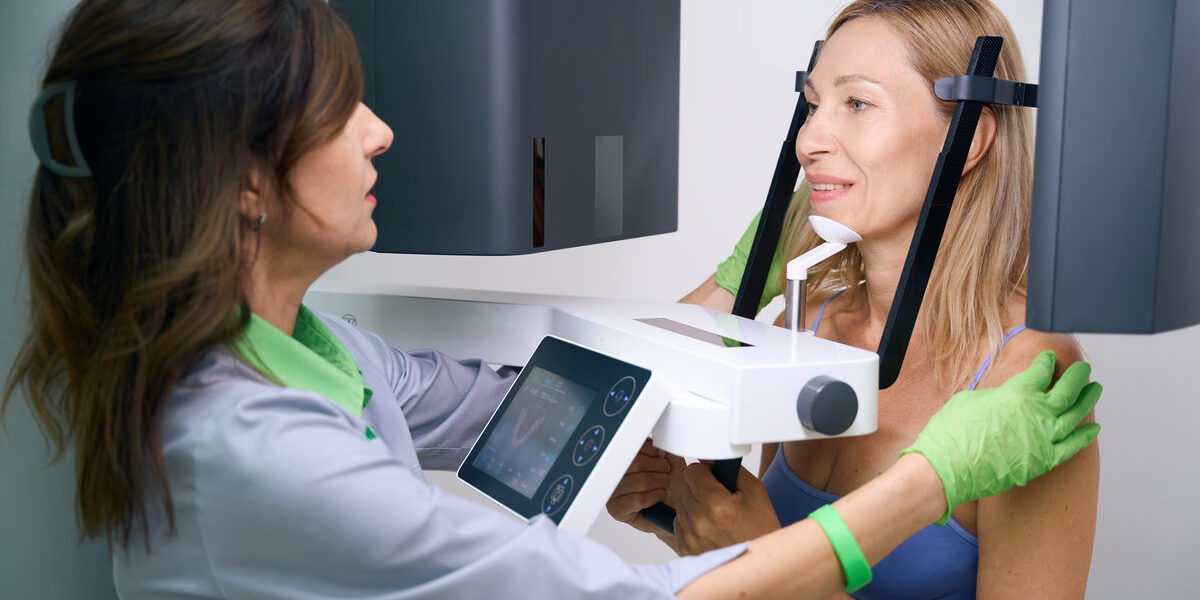
Cone beam computed tomography, more commonly referred to as CBCT, is the primary form of three-dimensional imaging used in implant dentistry. Unlike traditional dental X-rays, CBCT produces a full volumetric scan of your jaw, teeth, bone tissue, and soft structures in a single pass. This gives your specialist a comprehensive view of the area being evaluated, including the exact position of the mandibular nerve, the height and width of available bone, and the proximity of your maxillary sinuses.
Several types of digital imaging tools serve distinct purposes throughout the diagnostic process:
- Digital X-rays Deliver detailed two-dimensional images of tooth structure and early bone changes.
- CBCT scans Produce three-dimensional images used for implant planning and computer-guided surgical preparation.
- Intraoral cameras Capture high-resolution, real-time images of oral tissue and existing conditions.
- Digital scanners Create precise 3D models that eliminate the need for traditional impressions.
Together, these tools allow our specialists to build a thorough picture of your oral anatomy before your treatment plan is finalized, supporting more informed decisions at every stage of your care.
Why Imaging Accuracy Matters for Implant Placement
The success of a dental implant depends directly on the precision of pre-surgical planning. CBCT scans reveal whether sufficient bone volume is present to support an implant or whether bone grafting should be considered before moving forward. This distinction is critical because placing an implant into inadequate bone leads to complications that are far more difficult to manage after the fact. According to the American Dental Association, a 2024 expert panel review published in the Journal of the American Dental Association reaffirmed that appropriate imaging practices provide the diagnostic clarity essential to safe, effective oral health care decisions.
Our 3D imaging services also support the creation of computer-aided surgical guides, which help ensure each implant is placed at the optimal position and angle for both function and aesthetics. This level of planning is especially valuable for complex restorations like All-on-4 implants, where multiple implants must be positioned across the arch with exacting precision. Simulating placement digitally before the actual procedure reduces the margin for error and leads to more predictable, long-term results for patients.
What to Expect During Your Imaging Appointment
Digital imaging is a comfortable, efficient part of your pre-treatment evaluation. Digital sensors are smaller and smoother than traditional film holders, reducing any discomfort during the imaging process. Scans are completed quickly, which means less time spent in awkward positions during your visit.
Images appear on screen within seconds, allowing your specialist to review findings with you immediately and begin discussing your treatment options during the same appointment. This real-time exchange supports more productive consultations, giving you a clear understanding of your current oral health status and the recommended treatment. Digital records are also easily shared with your referring provider, ensuring consistent communication throughout your care.
Contact Rockland Dental Specialists Today
Dr. Shalom Mintz, Dr. David Peto, and our full team at Rockland Dental Specialists bring over 24 years of combined experience in periodontics, implants, and endodontics to every patient we see. Our advanced imaging capabilities, high-end office environment designed for your comfort, and commitment to personalized care reflect the standard we hold ourselves to for every case. Read through our patient testimonials to hear directly from patients who have experienced our approach to specialized dental care.
To schedule a consultation and get started on your implant treatment, contact our office today and let our team evaluate your needs with the thoroughness and precision your care deserves.